Media Release | June 12, 2013
Researchers at the University of British Columbia have developed a new magnetic resonance imaging (MRI) technique that detects the telltale signs of multiple sclerosis in finer detail than ever before – providing a more powerful tool for evaluating new treatments.

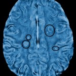
A frequency-based MRI image of an MS patient shows changes in tissue structure.
Download Full Size Image
Download Full Size Image
The technique analyzes the frequency of electro-magnetic waves collected by an MRI scanner, instead of the size of those waves. Although analyzing the number of waves per second had long been considered a more sensitive way of detecting changes in tissue structure, the math needed to create usable images had proved daunting.
Multiple sclerosis (MS) occurs when a person’s immune cells attack the protective insulation, known as myelin, that surrounds nerve fibres. The breakdown of myelin impedes the electrical signals transmitted between neurons, leading to a range of symptoms, including numbness or weakness, vision loss, tremors, dizziness and fatigue.
..
USE OUR SHARE LINKS at the top of this page – to provide this article to others
REMAIN up to date with MS News and Education
Visit our MS Learning Channel on YouTube: http://www.youtube.com/msviewsandnews


