In a study that used two colors to distinguish the cell types in a mouse model of multiple sclerosis, invading macrophages appear to be stripping myelin from axons, while resident microglia are virtually shut down
In a study that used two colors to distinguish the cell types in a mouse model of multiple sclerosis, invading macrophages appear to be stripping myelin from axons, while resident microglia are virtually shut down
CAROL CRUZAN MORTON
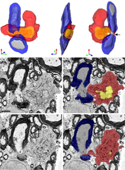
These scanning electron microscope images and three-dimensional reconstructions show a monocyte (red cytosol, yellow nucleus) next to a node of Ranvier from a mouse spinal cord, with two of the cell’s microvilli (black and white arrows) between the myelin (blue) and axon (gray). ©Yamasaki et al., 2014, Originally published in J. Exp. Med. doi: 10.1084/jem.20132477.
When a mob converges on a scene of impending destruction, it’s often hard to tell which individuals cause trouble and which ones may be innocent bystanders or even potential heroes. A new study provides the clearest picture yet that look-alike innate immune cells crowding around doomed myelin have distinct roles in disease.
In two types of images taken on day one of experimental autoimmune encephalomyelitis (EAE), an MS-like condition in mice, researchers caught invading monocytes from the blood red-handed in a direct attack on myelin. Meanwhile, the resident microglia of the central nervous system (CNS), which had also rushed to the scene, appeared to be nearly shut down. These two distinct groups of cells are both unhelpfully called macrophages and have been virtuallyindistinguishable in an inflamed CNS until recently.
“It’s a fantastic advance,” said Dwight Bergles, Ph.D., a neuroscientist at Johns Hopkins University, who was not involved in the study. “This study took advantage of genetics to paint the different populations of cells with different colors and unambiguously discriminate the different characteristics. It suggests they have different roles in disease. This is the beginning of a new series of studies.”
The findings were published in a cover article in the July 28 Journal of Experimental Medicine, by a team in the lab of Richard Ransohoff, M.D., a neurologist at the Cleveland Clinic in Ohio (Yamasaki et al., 2014).
“This paper sets a new standard for further studies in the field,” wrote Michael Heneka, M.D., a neurologist at the University of Bonn in Germany, in a commentary that accompanied the paper (Heneka, 2014). “For the first time, MDMs [monocyte-derived macrophages] and MiDMs [microglia-derived macrophages] have been clearly differentiated and the morphological relationship to axoglial structures has been analyzed.”
~~~~~~~~~~~~~~~~~~~~
Keep CURRENT and up to date, with MS News and Information
Sign-up for emails
.
WATCH OUR MS EDUCATIONAL VIDEOS by Topic,
found here: www.youtube.com/msviewsandnews
.
Visit our MS Learning Channel on YouTube: http://www.youtube.com/msviewsandnews
