February 11, 2016 /
FEBRUARY 10, 2016
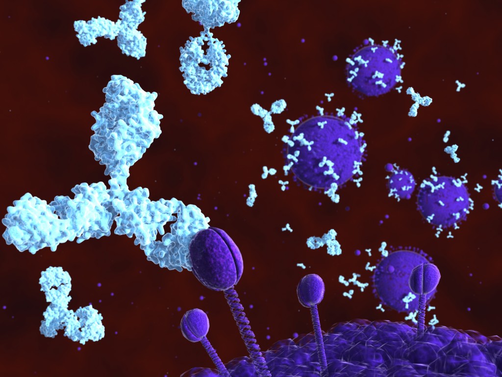
Brigham and Women’s Hospital researchers reported that antibodies directed at lipids are associated with magnetic resonance imaging (MRI) measures of brain degeneration in patients with multiple sclerosis (MS), and may potentially serve as biomarkers for monitoring disease status.
While the hyperintense brain lesions detected by MRI are crucial for diagnosis and therapeutic monitoring, they have proven to be of little value in predicting disease progression. Measurements of brain atrophy might associate better with clinical status. Earlier studies have shown that atrophy in the brain’s gray matter is more closely linked to disease status than atrophy in its white matter, and that compartment-specific measures need to be used when analyzing disease progression.
In the study “Serum lipid antibodies are associated with cerebral tissue damage in multiple sclerosis,“ researchers explored the possible relationships between immune factors, measured by an antigen array, and brain changes measured by MRI.
The research team, led by Rohit Bakshi, recruited 21 patients with relapsing-remitting MS (RRMS), and measured 420 different antigens present in serum samples taken from them. The patients also underwent MRI scans to assess the gray matter and white matter regions, as well as total brain tissue volume. Finally, researchers investigated lesion load based on the MRI scans.
READ MORE
~~~~~~~~~~~~~~~~~~~~~~~~~~~~~~~~~~~~~
MS Views and News
helps to provide educational information for persons affected by MS
Keep current with Multiple Sclerosis news and information
by opting-in to our website: click here – thank you
.===================================
Visit our MS Learning Channel on YouTube: http://www.youtube.com/msviewsandnews
Stay informed with MS news and information - Sign-up here
For MS patients, caregivers or clinicians, Care to chat about MS? Join Our online COMMUNITY CHAT

